This web page was produced as an assignment for Genetics 564, an undergraduate course at UW-Madison.
Background
Alzheimer's Disease, a common percursor to dementia, is a worldwide neurodegenerative disease of the brain, currently found in roughly 5 million people in the United States alone, 1 in 64 individuals. Symptoms of Alzheimer's Disease include, but are not limited to: short and long-term memory loss, speech impediment, and disorientation.
While there can be external causes for Alzheimer's, such as cranial damage and depression, studies indicate that roughly 70% of all diagnoses are due to genetic inheritance [6]. Individuals with Alzheimer's experience drastic reduction of the cerebral cortex and hippocampus, and an enlargement of cranial ventricles--leading to the many neurodegenerative symptoms observed with Alzheimer's Disease.
While there can be external causes for Alzheimer's, such as cranial damage and depression, studies indicate that roughly 70% of all diagnoses are due to genetic inheritance [6]. Individuals with Alzheimer's experience drastic reduction of the cerebral cortex and hippocampus, and an enlargement of cranial ventricles--leading to the many neurodegenerative symptoms observed with Alzheimer's Disease.
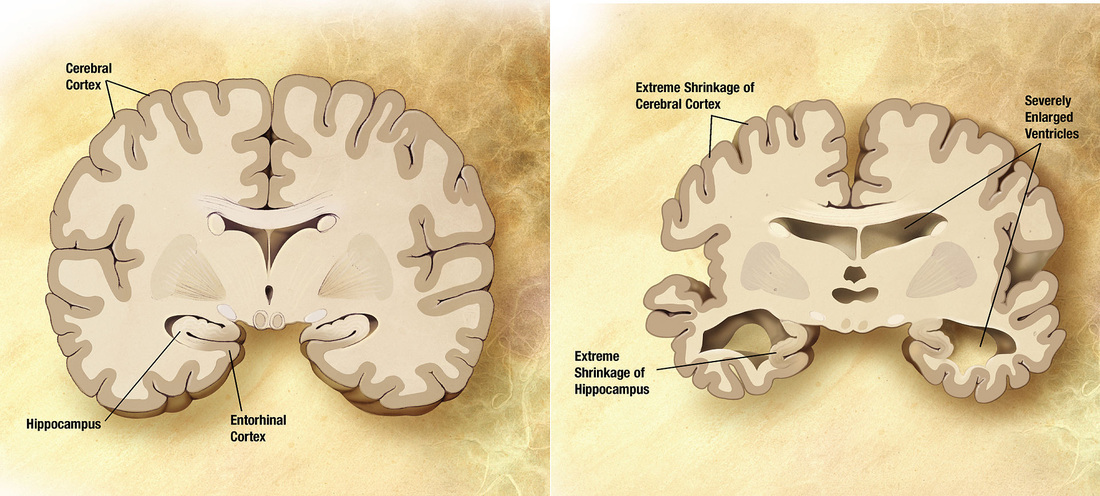
Figure 1. Brain structure between individuals with and without Alzheimer's. On the left is an individual without Alzheimer's, while the individual on the right has a progressive stage of Alzheimer's. Size of the cerebral cortex and hippocampus have decreased in those experiencing Alzheimer's Disease while the ventricles present have been enlarged.
Further illustrated in the video below, once Alzheimer's Disease has begun to progress, a build up of amyloid precursor protein leads to the production of senile plaques and neurofibrillary tangles in the brain's nervous system disrupt communication between neurons, causing the neurodegenerative symptoms observed in Alzheimer's. Similarly, disruption in cell communication causes progressive cell apoptosis, or death--leading to the decrease in brain function and size. As a result, various stages of Alzheimer's disease can be observed as follows:
1) Short term memory loss
2) Loss of coherent, logical thoughts
3) Erratic mood swings
4) Paranoia and hallucinations
5) Long term memory loss
6) Degradation of the brain
1) Short term memory loss
2) Loss of coherent, logical thoughts
3) Erratic mood swings
4) Paranoia and hallucinations
5) Long term memory loss
6) Degradation of the brain
Video 1. YouTube 4.3.2014. What is Alzheimer's Disease? Retrieved here.
While still a disease affecting many individuals in both the United States and on a global scale, there are several treatments proposed to negate, and eventually reverse the effects presented by Alzheimer's. The first option is an acetylcholine therapy treatment, which has been proven to stimulate brain activity in those diagnosed with Alzheimer's. By increasing brain function, neurons functions are exercised, reducing the amount of senile plaques formed and cell deaths.
The second option of treatment is a form of plaque vaccination. By directly reducing the amount of plaques formed through Alzheimer's in the nervous system, symptoms of Alzheimer's can be slowed and eliminated--preventing the stages of Alzheimer's from occurring before they begin.
Though no absolute cure for Alzheimer's has been developed, the world is currently devoted to isolating an Alzheimer's prevention technique. In doing so, it is important to understand the genetic involvement in Alzheimer's disease, namely Presenilin 1. Both the PSEN1 gene and PSEN1 protein are directly involved in developing Alzheimer's, outlined below.
PSEN1 Overview
|
PSEN1 Gene Location
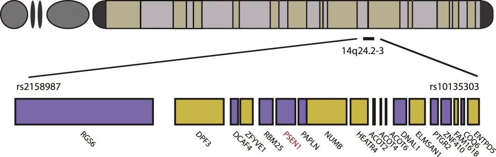
Figure 2. PSEN1 gene location in humans. Found in a small domain of chromosome 14, the complexity of the PSEN1 gene is illustrated.
|
PSEN1, a gene found in the human genome, is a crucial precursor to Alzheimer's Disease. Encoding for Presenilin I, a major protein component utilized in the presenilin complex, PSEN1 has been traced to development of Alzheimer's in individuals due to its involvement in producing the amyloid precursor protein (APP). Over-production of APP is linked to the development of Alzheimer's, as the toxicity of APP leads to the involved neurodegradation.
Found on human chromosome 14, PSEN1 can be traced to a series of homologs and model organisms. As such, model organisms can be used to further study and understand Alzheimer's disease. |
In such studies, it was discovered that a series of loss-of-function mutations lead to the increased production of APP through PSEN1 [1]. The mutation causes the PSEN1 gene to over-produce APP, increasing the cell toxicity, which in turn produces the neurological degradation depicted in figure 1. Similarly, the highly conserved regions of the PSEN1 gene indicate the usefulness of both homologs and model organisms when studying the gene.
PSEN1 Protein
|
PSEN1 protein, one of four components of the presenilin complex, has been discovered to directly mediate the production of gamma-secretase--a protein directly involved in further APP production [7]. As such, PSEN1 can be attributed as a direct causation of developing Alzheimer's disease. However, current studies indicate that when gamma-secretase is inhibited, APP is still over-produced in individuals with Alzheimer's [8].
Being a highly conserved protein, as depicted in Figure 3, there are several model organisms that can be utilized to test developing cases of Alzheimer's including Xenopus laevis and Drosophila melanogaster [5]. In utilizing such models, future studies on PSEN1 protein involvement can be studied. In addition, PSEN1 protein involvement with gamma-secretase has been found in several homologs and model organisms, observed in Figure 3 [2]. Because of this, model organisms can be utilized to test potential Alzheimer's Disease treatments revolved around gamma-secretase. Because gamma-secretase has been identified as a direct precursor to producing amyloid proteins, PSEN1 proves to be an important focus on current Alzheimer's research [3]. |
PSEN1 Protein Complex
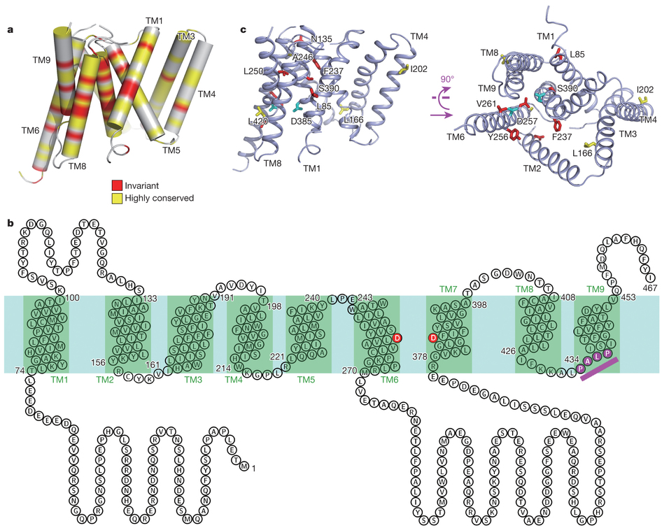
Figure 3. PSEN1 protein complex. Homology of human presenilin 1 to Xenopus laevis PS1 and Drosophila melanogaster PS1 indicates highly conserved protein structure in various model organisms.
|
Global Prevalence
Affecting five million individuals in the United States alone, the incidence of Alzheimer's has seemingly increased over the years--mainly due to the advancements in Alzheimer's diagnosis. While initial Alzheimer's research began early in 1901, it took 114 years to obtain the medical advancements gained today. With Alzheimer prevention techniques of acetylcholine treatment and plaque vaccination advancing, there is an increased global stance on Alzheimer's prevention. Affecting 44.4 million people globally, or 1 every 150 people worldwide, on a scale that continuously increases indicates just how important future Alzheimer's Disease experiments are [9]. Through further understandings of both the PSEN1 gene and PSEN1 protein, Alzheimer's Disease could be reduced and potentially one day eliminated in the global picture.
Regarding global prevalence, studies indicate that Alzheimer's Disease is most commonly found in Western Europe, while least identified in Sub-Saharan Africa [10]. Knowing this, mutations of both the PSEN1 gene and PSEN1 protein can be more readily traced to certain regions, useful in accurate identification of where these mutations initially occurred, and crucial to future Alzheimer's Disease and presenilin 1 studies.
Regarding global prevalence, studies indicate that Alzheimer's Disease is most commonly found in Western Europe, while least identified in Sub-Saharan Africa [10]. Knowing this, mutations of both the PSEN1 gene and PSEN1 protein can be more readily traced to certain regions, useful in accurate identification of where these mutations initially occurred, and crucial to future Alzheimer's Disease and presenilin 1 studies.
REFERENCES
YouTube video: https://www.youtube.com/watch?v=yJXTXN4xrI8
Figure 1. "Alzheimer's disease brain comparison" by derivative work: Garrondo (talk)SEVERESLICE_HIGH.JPG: ADEAR: "Alzheimer's Disease Education and Referral Center, a service of the National Institute on Aging."PRECLINICALSLICE_HIGH.JPG: ADEAR: "Alzheimer's Disease Education and Referral Center, a service of the National Institute on Aging." - SEVERESLICE_HIGH.JPGPRECLINICALSLICE_HIGH.JPG. Licensed under Public Domain via Wikimedia Commons - http://commons.wikimedia.org/wiki/File:Alzheimer%27s_disease_brain_comparison.jpg#mediaviewer/File:Alzheimer%27s_disease_brain_comparison.jpg
Figure 2. Cohen, J. (2013). Origin of Alzheimer's Gene Mutation Discovered. Obtained from http://www.news.ucsb.edu.
Figure 3. Li, X., Dang, S., Yan, C., Gong, X., Wang, J., Shi, Y. (2013). Structure of a presenilin family intramembrane aspartate protease. Nature, 493, 56-61.
[1] De Strooper, B. (2007). Loss-of-function presenilin mutations in Alzheimer disease. Talking Point on the role of presenilin mutations in Alzheimer disease.EMBO Reports, 8(2), 141–146. doi:10.1038/sj.embor.7400897
[2] Wolfe, M. S. (2013). Toward the structure of presenilin/γ-secretase and homologs. Biochimica et Biophysica Acta, 1828(12), 2886–2897. doi:10.1016/j.bbamem.2013.04.015
[3] Barnwell, E., Padmaraju, V., Baranello, R., Pacheco-Quinto, J., Crosson, C., Ablonczy, Z., … Sambamurti, K. (2014). Evidence of a Novel Mechanism for Partial γ-Secretase Inhibition Induced Paradoxical Increase in Secreted Amyloid β Protein. PLoS ONE, 9(3), e91531. doi:10.1371/journal.pone.0091531
[4] Cohen, J. (2013). Origin of Alzheimer's Gene Mutation Discovered. Obtained 02/16/2015 from http://www.news.ucsb.edu.
[5] Li, X., Dang, S., Yan, C., Gong, X., Wang, J., Shi, Y. (2013). Structure of a presenilin family intramembrane aspartate protease. Nature, 493, 56-61.
[6] Burns, A; Iliffe, S (5 February 2009). "Alzheimer's disease.". BMJ (Clinical research ed.) 338: b158.doi:10.1136/bmj.b158. PMID 19196745.
[7] St George-Hyslop P, Fraser P (January 2012). "Assembly of the presenilin γ-/ε-secretase complex". J. Neurochem. 120 Suppl 1: 84–8. doi:10.1111/j.1471-4159.2011.07505.x. PMID 22122073.
[8] Selkoe D (1994). "Cell biology of the amyloid beta-protein precursor and the mechanism of Alzheimer's disease". Annu. Rev. Cell Biol. 10: 373–403.doi:10.1146/annurev.cb.10.110194.002105.PMID 7888181.
[9] Dimentia Statistics. Obtained from http://www.alz.co.uk/research/statistics.
[10] Alzheimer's Statistics. Obtained from http://www.alzheimers.net/resources/alzheimers-statistics/.
